Trichinoscopes
Trichinoscopes are microscopes that were specifically designed to diagnose trichinella, the presence of the parasite Trichinella spiralis in muscle tissue samples of animals that are the basis of food for humans.
Fig.1. Preparation seen under the microscope of pig striated muscle with trichinosis. Photograph of the author.
Trichinosis
Also known as trichinellosis, it is a parasitic disease caused by eating undercooked meat and containing cysts (larvae or immature worms) of Trichinella spiralis. This parasite can be found in the meat of animals such as pig, horse, wild boar, deer and rat.
It is a common infection in the world and it was very frequent in the past. When a person eats meat from an infected animal, trichina cysts, present in the meat, hatch in the intestines and grow into adult nematodes. Nematodes produce other worms that migrate through the intestinal wall into the bloodstream. The worms invade muscle tissues, which include the heart and diaphragm. They can also affect the lungs and brain. Cysts stay alive for years. Trichinosis symptoms include: abdominal discomfort, spasms, diarrhea, facial edema, fever, and especially muscle aches.
The signs, symptoms and their severity vary according to the number of larvae that were consumed with the infected meat. It is a self-limited disease but it can present complications.
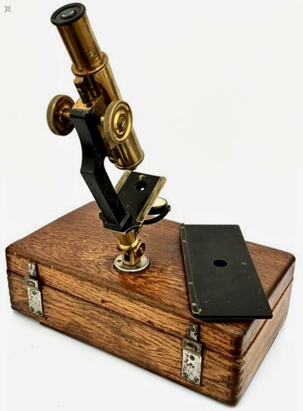
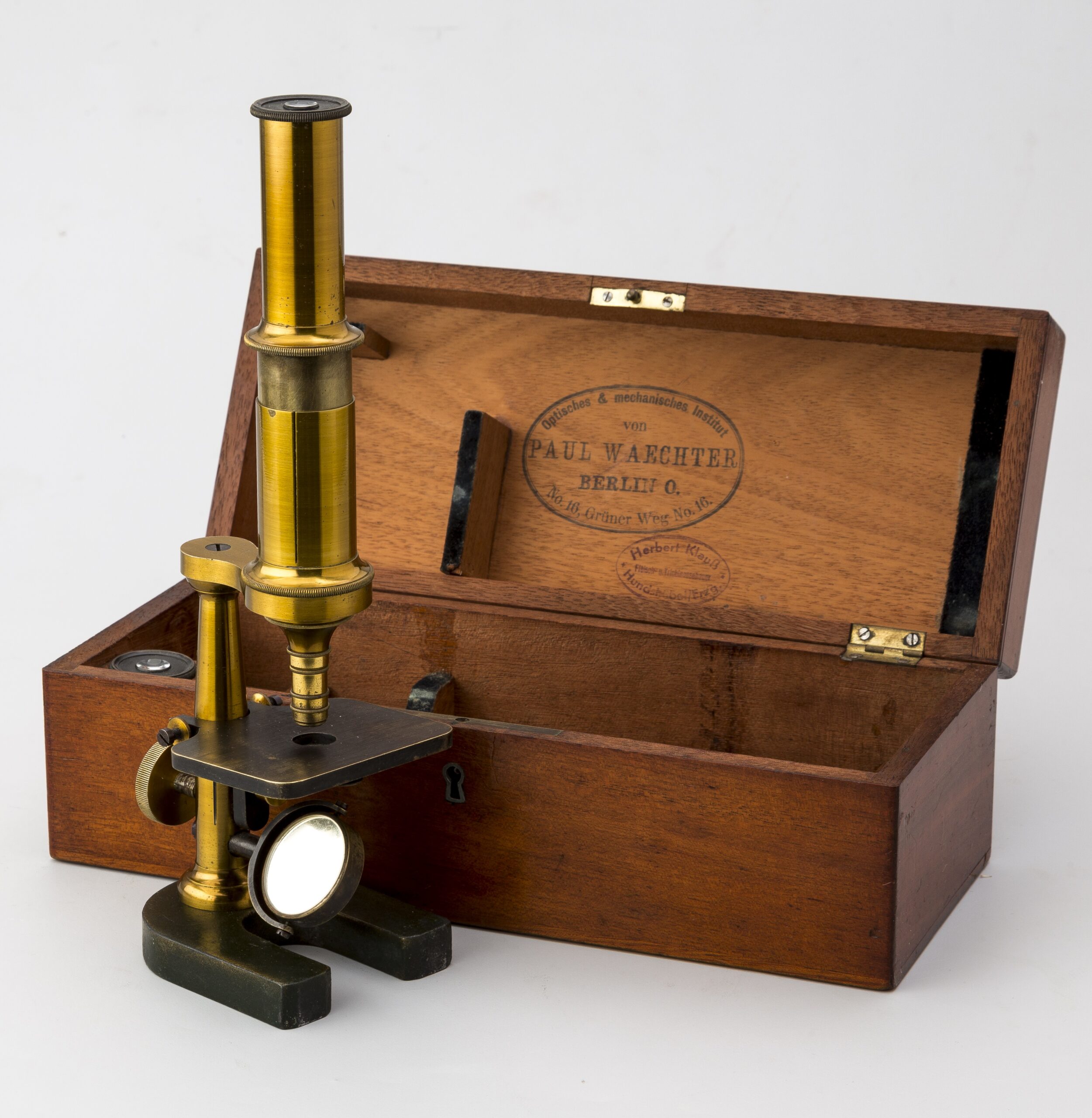
20a. Microscopio triquinoscopio de Paul Waechter. Alemania c.1920.
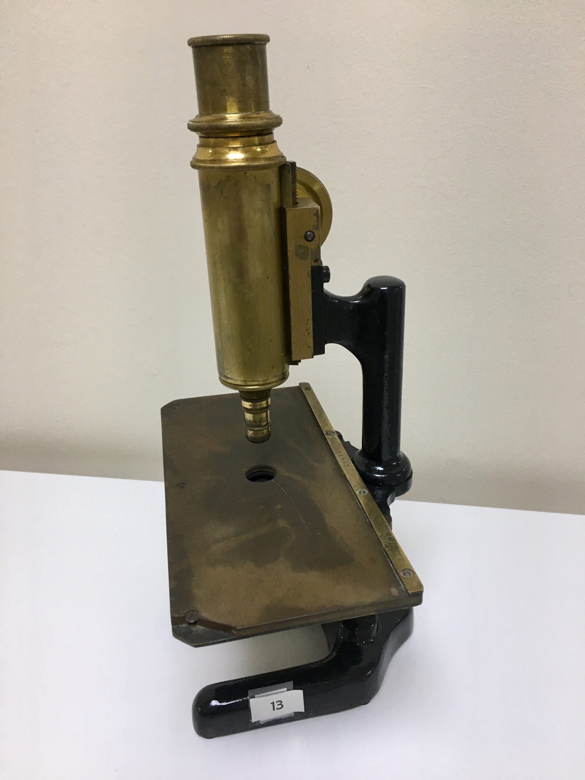
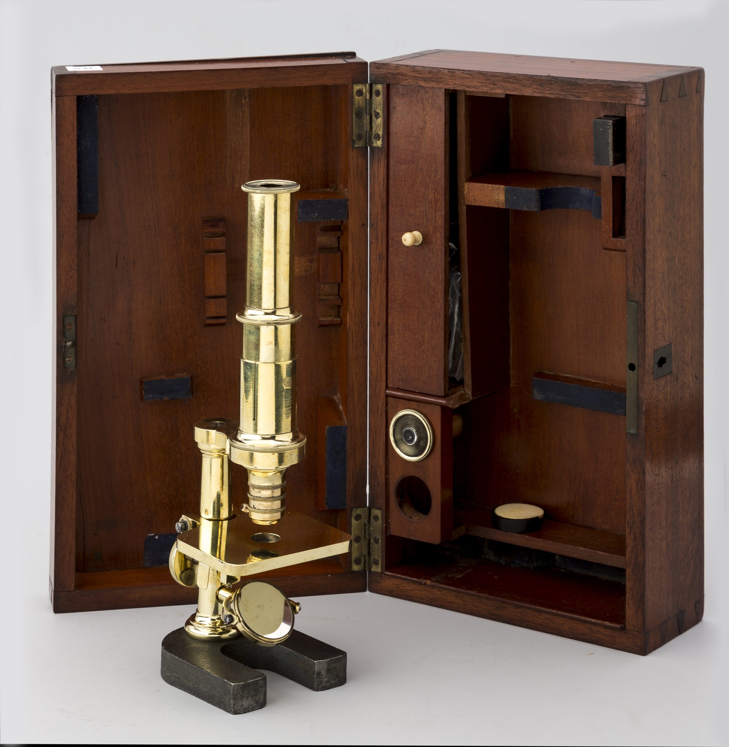
43a.bis. Triquinoscopio portátil. H. Hauptner. Berlín – Solingen. c.1870.
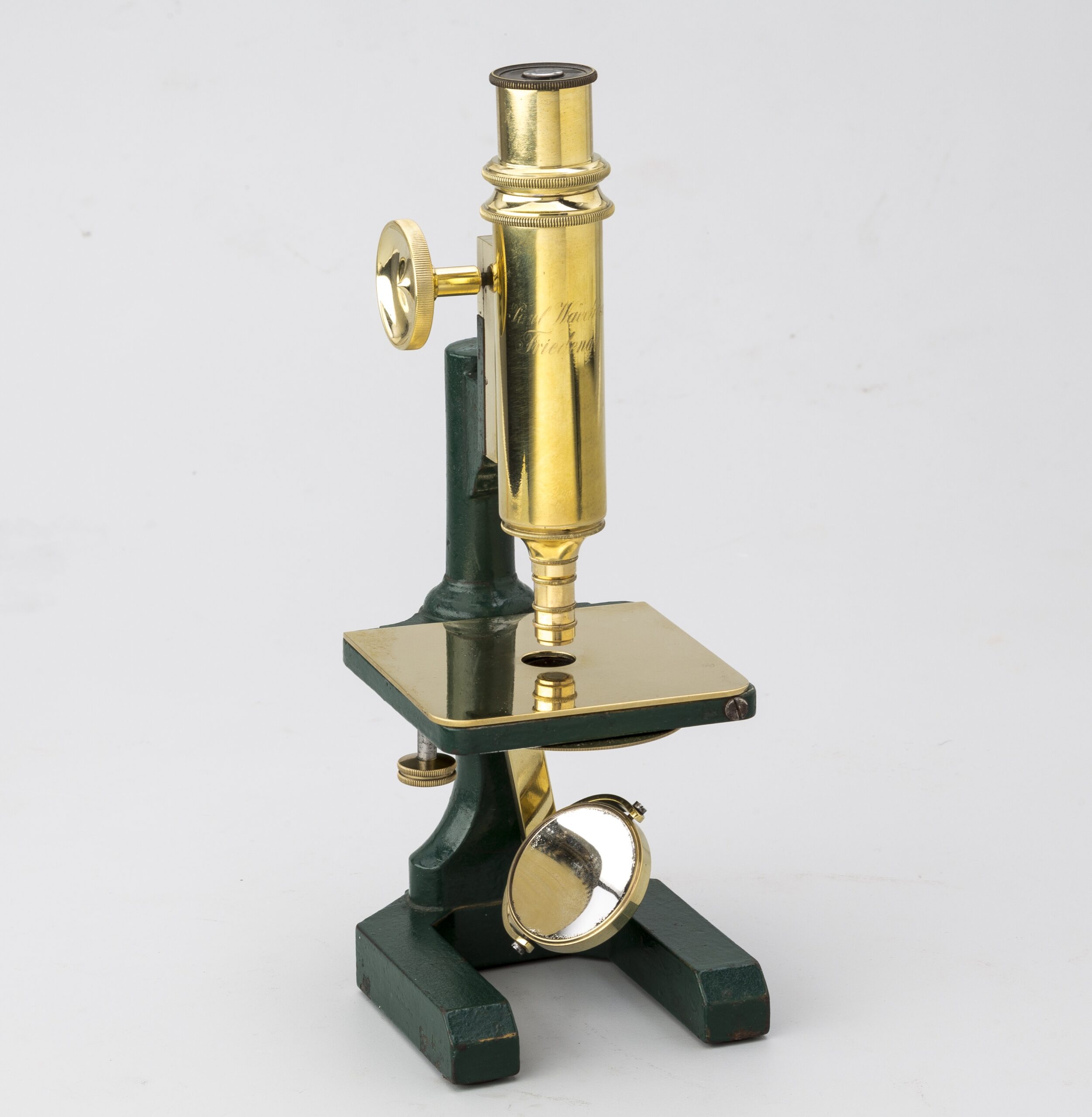
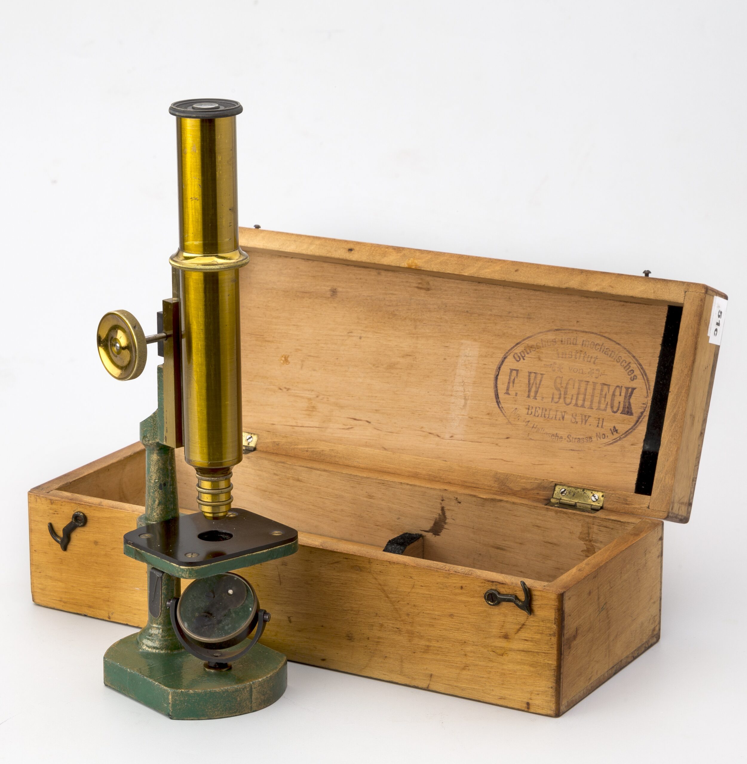
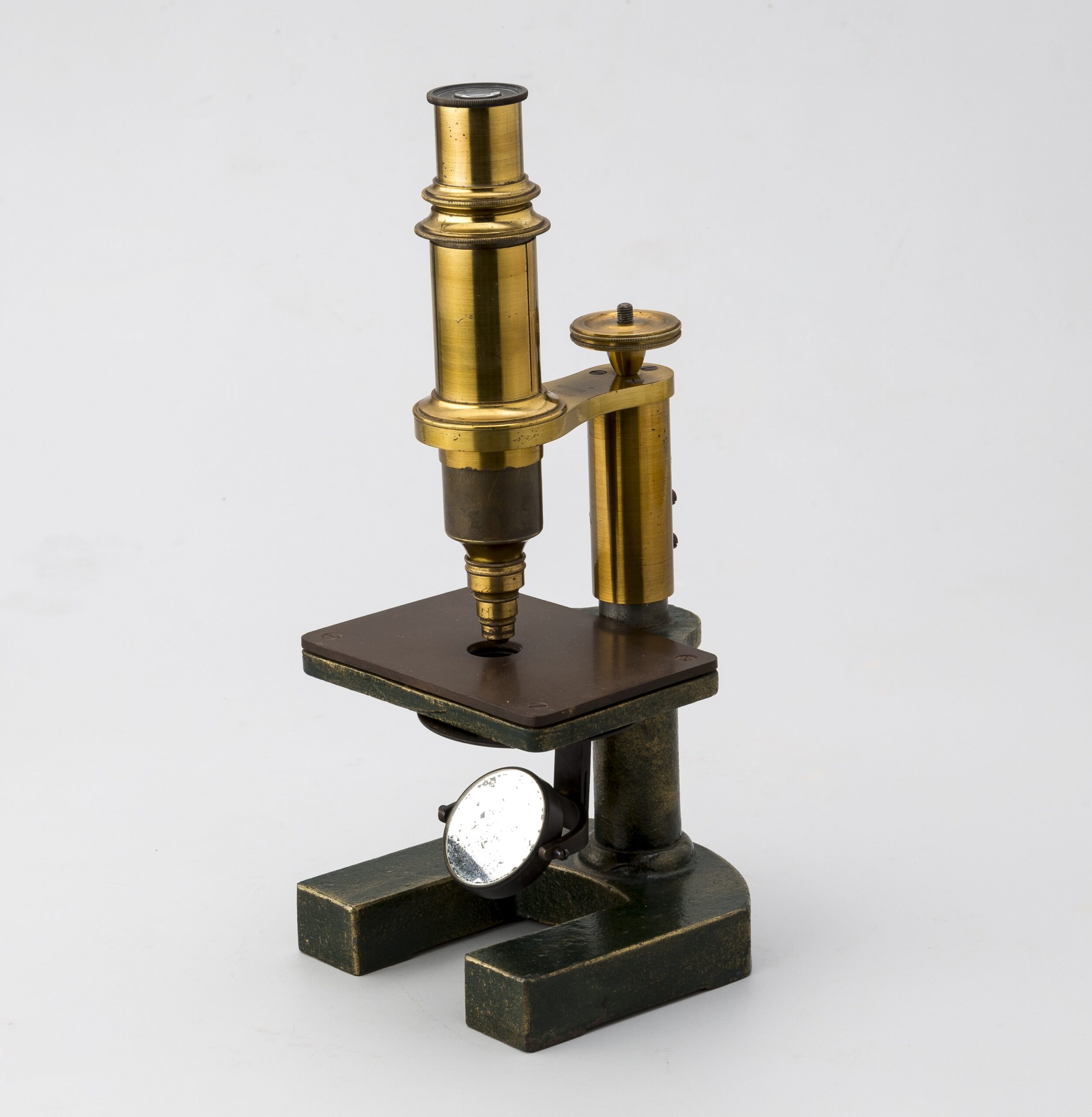
51d. Triquinoscopio. Waechter P.Friedenau. c.1875.
51e. Microscopio monocular Waechter P. Berlín. Modelo Stativ IV.
51f. Microscopio monocular Waechter P. Berlín. Modelo Stativ V
51g.Triquinoscopio P. Thate. Berlin. c.1865.